Submitted by admin on Sat, 09/20/2008 - 21:10
tissue_organ_import:
heart
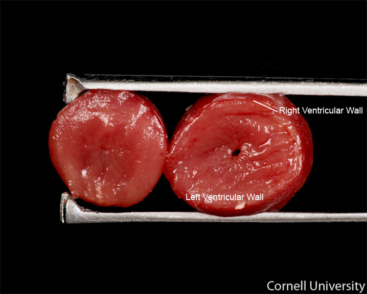
Clinical description:
The bottom 1/3 of the apex of the heart has been removed. This view allows the thickness of the ventricular walls and septum to be assessed. The right ventricular free wall should be approximately 1/3 to 1/2 the thickness of the intraventricular septum and the left ventricular free wall. In the normal heart, the lumina of the ventricles should be small and there should be negligible space between the walls of the ventriculae and the septum. In cardiac diseases, such as pulmonary hypertension or congenital defects, this space will be widened.
Record number:
10279
Case number:
Unknown
Age:
6 weeks
Breed:
White Leghorn
Clinical form:
Unknown
Infection type:
Unknown
Housing/mgmnt type:
Select One
Priority:
1
Rights:
© Cornell University
Etiology:
Tissues and organs:
Asset type:
Species:
Image: