Submitted by admin on Sat, 09/20/2008 - 21:10
tissue_organ_import:
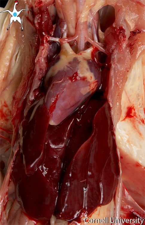
heart
Clinical description:
With the bird in dorsal recumbency, the walls of the right atrium and ventricle are positioned on top. After death, the atria often expand with blood and can appear quite enlarged. The great vessels can be seen entering the base of the heart. The heart muscle should be reddish-brown in color. A variable amount of fat will be present in the coronary grooves. If the bird is emaciated, this fat may be absent or have undergone serous atrophy, resulting in a gelatinous, wet appearance. Look for any external lesions on the epicardial surface of the heart or on the surrounding fat.
Record number:
10249
Case number:
Unknown
Age:
6 weeks
Breed:
White Leghorn
Clinical form:
Unknown
Infection type:
Unknown
Housing/mgmnt type:
Select One
Priority:
1
Rights:
© Cornell University
Etiology:
Tissues and organs:
Asset type:
Species:
Image: