Submitted by admin on Sat, 09/20/2008 - 21:10
tissue_organ_import:
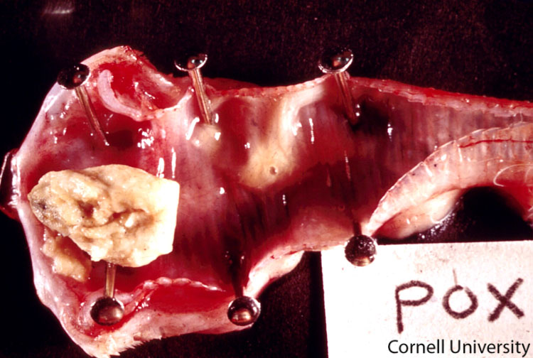
trachea
Morphologic diagnosis:
Trachea: Diffuse necrosuppurative tracheitis with locally extensive diptheretic membrane.
Clinical description:
This image shows a typical diphtheritic lesion caused by avian pox. The fibrino-necrotic proliferative lesions form on mucous membranes. These lesions may develop in the oral cavity, tongue, esophagus, or upper trachea. If the lesions are located in the lower respiratory tract (e.g. the syrinx), they can compromise breathing, resulting in dyspnea.
Pathologic description:
The trachea has been partially opened to reveal the mucosal surface which is diffusely red. Within the lumen there is a focal accumulation of wet, pale tan, mucoid material along with a partially occlusive plug composed of dry, friable, yellow tan material.
Record number:
7669
Case number:
Unknown
Clinical form:
Unknown
Infection type:
Unknown
Housing/mgmnt type:
Select One
Priority:
1
Image source URL:
http://cidc.library.cornell.edu/vet_avian/images/POX Adjusted/POX-050A.jpg
Etiology:
Exam findings:
Tissues and organs:
Asset type:
Species:
Image: