
Submitted by admin on Tue, 08/26/2008 - 15:40
tissue_organ_import:
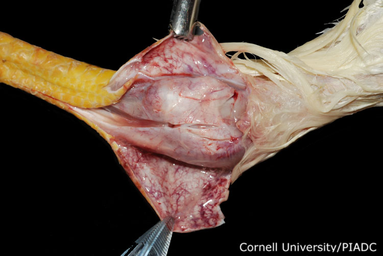
Intertarsal (hock) joint: subcutaneous edema.
Morphologic diagnosis:
Subcutaneous and periarticular tissue: Acute edema and congestion.
Clinical description:
This image was taken 3 days post experimental inoculation with highly pathogenic avian influenza. An incision has been made over the swollen hock joint, revealing a subcutaneous accumulation of serohemorrhagic exudate, consistent with edema.
Pathologic description:
The skin overlying the swollen joint has been peeled back to reveal the subcutaneous tissue and portions of the joint capsule. The tissue is slightly wet, and the blood vessels are prominent. Near the left side of the photo, there is a short linear incision into the joint capsule and the tendon attachment. This incision reveals the accumulation of a small amount of yellow tinged fluid in the tissue.
Record number:
20923
Case number:
5048
Age:
16 weeks
Breed:
White Leghorn SPF
Clinical form:
Acute
Infection type:
Experimental
History:
The photograph was taken 3 days post inoculation. The bird was experimentally inoculated with highly pathogenic avian influenza virus on 3/2/08 at Plum Island Animal Disease Center. The inoculation was performed in the caudal thoracic air sac with strain A/CK/PA/469/3-84/H5N2, using 0.25ml.
Housing/mgmnt type:
Select One
Priority:
1
Etiology:
Exam findings:
Tissues and organs:
Asset type:
Species:
Image:
- Log in to post comments