
Submitted by admin on Tue, 08/26/2008 - 15:40
Clinical Signs:
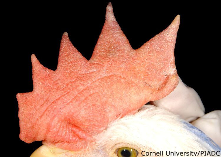
Necrosis, cyanosis (comb)
Morphologic diagnosis:
Comb: Mild multifocal necrosis and cyanosis
Clinical description:
This image was taken 2 days post experimental inoculation with highly pathogenic avian influenza. The tips of the comb are beginning to show signs of early necrosis and cyanosis. These lesions form as microthrombi develop within the dermal vessels of the nonfeathered skin. Thrombi lead to vasculitis, perivascular edema, subcutaneous edema, and finally necrosis of the capillary endothelium. The degree of edema, hemorrhages, and necrosis present will depend on the stage of this process.
Pathologic description:
The tips of the comb are dry, shrunken and slightly blue. The changes are particularly evident on the second tip from the right. In this area, the skin is slightly purple and the tip is constricted, slightly brown and covered by several petechia. The outer-most extent of the tip is brown and dry.
Record number:
20733
Case number:
5048
Age:
6 weeks
Breed:
White Leghorn SPF
Clinical form:
Acute
Infection type:
Experimental
History:
The photograph was taken 2 days post inoculation. The bird was experimentally inoculated with highly pathogenic avian influenza virus on 3/2/08 at Plum Island Animal Disease Center. The inoculation was performed in the caudal thoracic air sac with strain A/CK/PA/469/3-84/H5N2, using 0.25ml.
Zootechnical purpose:
Egg layers- white egg
Housing/mgmnt type:
Select One
Priority:
1
Rights:
© Cornell University
Etiology:
Exam findings:
Tissues and organs:
Asset type:
Species:
Image:
- Log in to post comments