
Submitted by admin on Tue, 08/26/2008 - 15:40
tissue_organ_import:
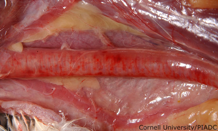
Trachea: hemorrhage and congestion.
Morphologic diagnosis:
Trachea: Diffuse hemorrhage and congestion
Clinical description:
This image was taken 2 days post experimental inoculation with highly pathogenic avian influenza. In HPAI, hemorrhagic tracheitis may be severe enough to be visible on the outer surface of the trachea.
Pathologic description:
The blood vessels on the adventitial surface of the trachea are congested and prominent. They are evident running parallel to the tracheal rings. The trachea is diffusely red and irregular red to purple foci can be seen throughout the tracheal wall.
Record number:
20791
Case number:
5005
Age:
70 weeks
Breed:
White Leghorn SPF
Clinical form:
Acute
Infection type:
Experimental
History:
The photograph was taken 2 days post inoculation. The bird was experimentally inoculated with highly pathogenic avian influenza virus on 3/2/08 at Plum Island Animal Disease Center. The inoculation was performed in the caudal thoracic air sac with strain A/CK/PA/469/3-84/H5N2, using 0.25ml.
Housing/mgmnt type:
Select One
Priority:
1
Rights:
© Cornell University
Etiology:
Exam findings:
Tissues and organs:
Asset type:
Species:
Image:
- Log in to post comments