
Submitted by admin on Tue, 08/26/2008 - 15:40
tissue_organ_import:
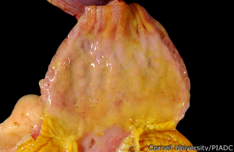
Proventriculus: hemorrhages and edema.
Morphologic diagnosis:
Proventriculus, ventriculus: Multifocal acute hemorrhages and diffuse edema
Clinical description:
This image was taken 2 days post experimental inoculation with highly pathogenic avian influenza. In HPAI, the glands of the proventriculus may have hemorrhagic lesions. As observed here, these hemorrhages tend to be concentrated at the junction between the proventriculus and the gizzard (ventriculus).
Pathologic description:
The mucosal surface and wall of the proventriculus is wet and glistening. Small pinpoint red foci are present within the proventricular glands. These hemorrhages are most dense at the junction of the proventriculus and the ventriculus (lower left of photo). A portion of the ventricular koilin has been peeled back (bottom center) to reveal hemorrhages in the subadjacent mucosa. Reddening of the mucosa at the esophageal/proventricular interface is also prominent.
Record number:
20786
Case number:
5005
Age:
70 weeks
Breed:
White Leghorn SPF
Clinical form:
Acute
Infection type:
Experimental
History:
The photograph was taken 2 days post inoculation. The bird was experimentally inoculated with highly pathogenic avian influenza virus on 3/2/08 at Plum Island Animal Disease Center. The inoculation was performed in the caudal thoracic air sac with strain A/CK/PA/469/3-84/H5N2, using 0.25ml.
Housing/mgmnt type:
Select One
Priority:
1
Rights:
© Cornell University
Etiology:
Tissues and organs:
Asset type:
Species:
Image:
- Log in to post comments